
Leica SP8 WL inverted confocal

Technology Focus
Confocal microscopes use a pinhole to reject out of focus light resulting in optical sectioning and an improved signal to noise ratio when compared to widefield fluorescence imaging. (See figure below – only in-focus light can pass through the pinhole to reach the detector).

Confocal microscopes are ideal for imaging multi-labelled fluorescent samples up to around 100 microns in depth.
Figure credit – https://www.edinst.com/

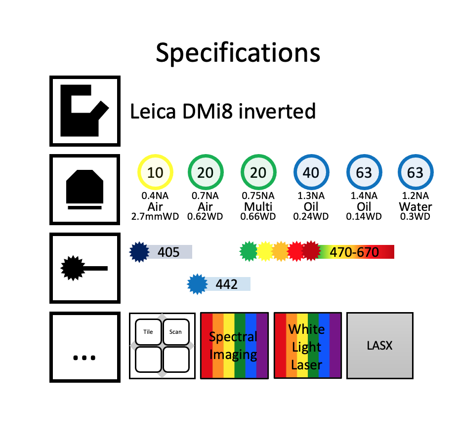
The White Light (WL) Laser ranges from 470nm to 670nm and 8 wavelengths can be simultaneously selected. This, together with the spectral detection, allows for easy optimization of multicolour imaging set-ups. The WL laser is pulsed giving the option of gating to remove reflections and other non fluorescent events from the images when used with the pulsed HyD detectors.
Training requests and equipment bookings are done through PPMS